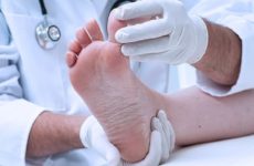
Numerous warts and other growths covering the face, hands and other parts of the body are common. Many people are accustomed to their presence on the skin, considering it a minor defect. In fact, the appearance of such seals reflects the state of human health.

Содержание:
Why are growths dangerous?
The habit of rubbing your eyes with your hands and exposure to ultraviolet rays can provoke an injury to the growth, followed by infection, which leads to the growth of the wart or its transformation into a malignant tumor.
Pathological seals of the skin interfere with normal vision, blinking and are located in close proximity to the eyeball, which can cause the development of neoplasms on the mucous membrane.
Varieties of growths
Depending on the etiology of the occurrence of compaction, there are:
- growths resulting from the penetration of an infectious agent into the body;
- enlargement of areas of the skin that are not associated with exposure to viruses or bacteria.
Formations of an infectious nature:
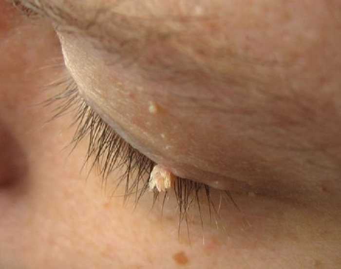
- papillomas are superficial growths that look like a small bag hanging on a thin stem or a small bulge on the surface of the eyelid. The color is identical to that of healthy skin. Papillomas can be single or plentifully cover the body;
- warts are neoplasms of various sizes. In shape, they are flat (small growths), ordinary (in the form of a brown or flesh-colored papule with a porous, rough surface) and finger-shaped (thick, elongated).
According to the principle of the probability of provoking the development of cancer, seals of viral etiology are classified:
- non-oncogenic group – there is no risk of developing a malignant tumor;
- oncogenic group with a low degree of risk ;
- oncogenic group with a high level of risk – infection with viruses of this type is manifested by the development of cancer in more than 40% of cases.
Benign seals, non-infectious nature:
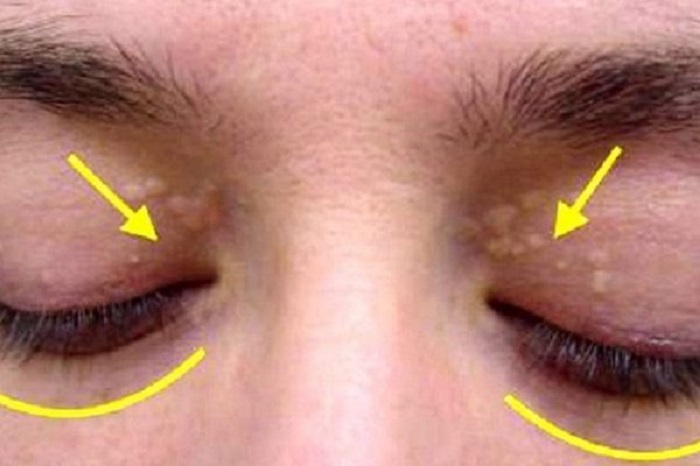
- syringomas are small (2-3 mm) round growths with a loose structure that occur as a result of thickening of the sweat glands. They have a yellowish tint, most often located on the lower eyelid;
- sebaceous gland hyperplasia h – round seals (from 2 mm to 10 mm), slightly protruding above the skin surface, with a granular surface and a depressed area in the center. The neoplasm has a light yellow tint;
- xanthelasmas – sac-like, characterized by an irregular shape, yellow-white color and a size of 2-10 mm. People with high levels of cholesterol in the blood are prone to the appearance of growths. Most often, xanthelasmas are located on the upper eyelid;
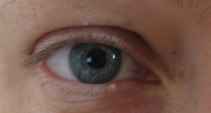
- milia – are formed due to blockage of the sebaceous glands;
- keratomas – flat warts of brown-yellow color, with a loose crust on the surface. Often occur in people who have been exposed to sunlight for a long time or because of the tendency of the stratum corneum of the epidermis to thicken.

Causes of occurrence on the lower or upper eyelid and around the eyes
The appearance of unwanted growths in the eye area occurs due to infection with the human papillomavirus.
The infection is transmitted by household means (through touch or when visiting public places – toilets, swimming pools, gyms), can pass from mother to newborn child.
The infectious agent is activated and provokes tissue growth under the influence of certain circumstances:
- hypothermia, living in cold climates;
- exposure to stress and frequent overwork;
- weakened immunity, beriberi, regular colds;
- prolonged exposure to ultraviolet rays;
- diseases of the genitourinary system, gastrointestinal tract;
- increased sweating;
- use of contraceptives based on hormones.
When should you see a doctor?
Diagnosis and treatment under the supervision of a physician is required in the following cases:
- wart bleeding due to trauma;
- discoloration of the skin growth, darkening;
- a sharp increase in the size of education in the eyelids.
Diagnosis of the disease
The method of diagnosing a build-up is determined by a dermatologist:
- colposcopy – the study of the epithelial tissues of the wart under a microscope;
- pap test – a cytological study of scraped biomaterial by staining with chemical reagents. The method serves to determine the presence of changes in cells;
- polymerase chain reaction – a method to determine the type of virus.
How to distinguish from other formations
- papillomas are located closer to the blood vessels than warts, so they bleed at the slightest injury;
- surface structure – warts have smooth edges and a rounded shape, papillomas are loose, hanging over the surface of the skin on a thin stalk;
- unlike moles, the density of the wart is much higher, but the pigmentation of the surface is less. Its main characteristic feature is the local thickening of the skin in the form of a hemisphere with a rough surface.
How to withdraw education?
Below we briefly consider the most effective methods for removing a wart and its treatment with drugs.
With the help of pharmaceuticals
Removal, possibly with combined bactericidal preparations based on phenol and tricresol:
- Ferezol – apply to the wart for 10-60 minutes (depending on the size of the seal), avoiding contact with neighboring skin areas. If you need to remove the papilloma – lubricate only the leg. Repeat the treatment one week after the dry crust falls off.
- Verrukacid – treat small growths once or 3-4 times larger growths using an applicator or a thin stick. Avoid contact with mucous membranes or adjacent integuments.
Folk remedies and recipes
lubricate the growth with fresh celandine juice 2 times a day, after treating the skin around the wart with vegetable oil. The neoplasm will disappear in a few days:
- within 5-7 days, apply 1 drop of vinegar essence to the seal;
- before going to bed, grind a piece of chalk and sprinkle the build-up, fix a piece of cloth on top, previously heated on the battery. Do not let the bandage get wet;
- collect yellow dandelion flowers, fill a small jar with them and fill with ethyl alcohol. Leave the solution for 10-14 days, placing the container in a dark place, then strain and lubricate the wart 4-5 times a day;
- pour the dry grass of lemon balm, nettle, plantain, dandelion and horsetail, taken in equal parts, into an enameled pan and fill with warm water. Put the dishes in a water bath and simmer for 10-15 minutes, then wrap the container with a blanket and leave for 3 hours. Use the collection for 7 days 3 times a day, 45 ml each (20-30 minutes before meals);
- lubrication with tea tree oil also helps the seal dry;
- Apply sour green apple juice 5 times a day until the wart disappears completely.

Hardware and surgical methods
The choice of a method for removing a wart on the eyelid is carried out by an ophthalmologist surgeon:
- electrocoagulation – the destruction of tissues by electric current at high temperature. A thin crust remains at the site of the wart, which must be lubricated with a special ointment for at least a week. The advantage of the procedure is the ability to control the depth of exposure, the disadvantage is damage to neighboring integuments, the duration of the healing period;
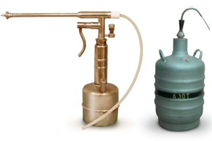
- cryodestruction – freezing of the growth with liquid nitrogen. The advantage of this type of intervention: no scars and scars. Disadvantages: danger and risk of injury due to the proximity of mucous tissues;
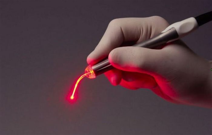
- laser – the operation is carried out by cutting out the seal and cauterizing its location with laser beams. Before the procedure, local anesthesia is performed. This is a safe and effective method, characterized by a rapid recovery of the health of the operated person;
- radio wave – a high-frequency radio signal supplied from the active electrode evaporates the intracellular fluid from the seal, after which it is excised and removed. Using the method allows you to minimize the side effects of the procedure and the risk of infection. It is also possible to conduct a histological examination of the tissues of the growth;
- surgical removal – used in case of large neoplasms. It occurs under local anesthesia with surgical instruments, by excising the body of the wart, followed by suturing and regular dressings. To eliminate seals on the eyelids, this method is used in rare cases.
Prevention and prognosis
The timeliness of contacting a doctor, strict adherence to the regimen of prescribed drugs and an integrated approach (simultaneous use of antiviral and immunostimulating agents) provide a favorable treatment prognosis.
Measures to prevent the appearance of warts:
- in order to avoid infection with papillomavirus, protect the skin of the eyelids from injuries: wounds, scratches;
- do not squeeze out the neoplasm, do not pull and do not try to get rid of it yourself;
- carefully follow the rules of personal hygiene;
- adhere to the principles of a balanced diet, give up bad habits;
- stay calm in stressful situations.
Pathological changes in the skin can occur anywhere on the body, but the eyelids are a particularly dangerous area.
The simple observance of preventive measures and seeking help from a doctor will eliminate the possibility of an unpleasant growth and its degeneration into a formidable, malignant neoplasm.


